Managing Pressure Ulcers: A Comprehensive Guide for Nurses and Therapists
Pressure ulcers, also known as bedsores or decubitus ulcers, represent a significant challenge throughout the world. For example, they often affect immobile or bedridden patients. As a result, this leads to pain, infection, and extended hospital stays. As nurses and therapists, your role involves prevention, early detection, and tailored interventions. This comprehensive guide provides practical strategies for managing pressure ulcers. Use it to optimize patient outcomes in hospitals, long-term care, or home health. Consequently, implementing these techniques can help to reduce incidence rates. You’ll also enhance quality of care in this common yet preventable condition.
Understanding Pressure Ulcers and Their Causes
Pressure ulcers develop when prolonged pressure on the skin restricts blood flow. Over time, this causes tissue damage. It typically occurs over bony prominences like the sacrum, heels, or hips. In addition, contributing factors include immobility and poor nutrition. Moisture from incontinence and shear forces during transfers also play a role. A pressure injury is localized damage to the skin and underlying soft tissue. It usually happens over a bony prominence or relates to a medical device. Before we discuss how to manage pressure ulcers, let’s look at the stages.
Stages of Pressure Ulcers: Overview
Stages range from Stage 1 to Stage 4. Additionally, categories include unstageable ulcers and deep tissue injuries. Nurses and therapists must recognize these stages. This ensures accurate assessment and appropriate management. As a result, early intervention can halt progression. It also avoids complications like osteomyelitis.
Stage 1: Early Skin Changes
Intact skin shows non-blanchable erythema (redness) in a localized area. This is usually over a bony prominence. The skin remains unbroken. However, the area may feel painful, firm, soft, warmer, or cooler than adjacent tissue. In darkly pigmented skin, blanching may not be visible. Color changes could appear as purple or blue hues instead of red. This early stage signals potential damage. Consequently, it requires immediate pressure relief to prevent progression.
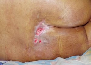
Stage 2: Partial-Thickness Damage
Partial-thickness loss of the dermis presents as a shallow open ulcer. It has a red-pink wound bed without slough or bruising. It may appear as an intact or open/ruptured blister filled with serum. The ulcer is superficial; it does not extend into subcutaneous tissue. But, it can be painful and prone to infection if not protected.
Stage 3: Full-Thickness Involvement
Full-thickness tissue loss makes subcutaneous fat visible. Bone, tendon, or muscle is not exposed. Slough may be present. But it does not obscure the depth of tissue loss. Undermining and tunneling are common. Additionally, the ulcer may extend into deeper layers. But, it stops short of underlying structures. This stage often needs debridement and advanced dressings.
Stage 4: Severe Tissue Loss
Full-thickness tissue loss exposes bone, tendon, or muscle. Slough or eschar may appear in parts of the wound bed. Undermining or tunneling is frequent. These ulcers can lead to osteomyelitis or sepsis if untreated. For this reason, these types of wounds necessitate multidisciplinary intervention.
Unstageable: Obscured Depth
Full-thickness tissue loss obscures the ulcer base completely. Slough (yellow, tan, gray, green, or brown) and/or eschar (tan, brown, or black) cover it. Remove enough slough or eschar to expose the wound bed. Only then can you determine the true depth and stage.
Deep Tissue Injury: Hidden Damage
A purple or maroon localized area shows discolored intact skin. Or it appears as a blood-filled blister. This stems from damage to underlying soft tissue due to pressure and/or shear. The area may feel painful, firm, mushy, boggy, warmer, or cooler than adjacent tissue. Evolution may include a thin blister over a dark wound bed. And, the wound may become covered by thin eschar.
Assessing Risk and Staging Ulcers
Effective management starts with risk assessment. Use tools like the Braden Scale. It evaluates sensory perception, moisture, activity, mobility, nutrition, and friction/shear. Conduct daily skin inspections. Document color changes, warmth, or induration. Stage ulcers accurately upon discovery. Measure size, depth, and exudate. Note undermining or tunneling. Reassess frequently to track changes. Involve the entire care team for accurate classification. Differentiate from other skin issues like moisture-associated damage.
Prevention Strategies in Daily Practice
Prevention is paramount. Begin with regular repositioning every two hours for bedbound patients. Use supportive surfaces like air-fluidized beds or foam mattresses. These redistribute pressure. Maintain skin hygiene with gentle cleansing and moisturizing. Avoid excessive friction during turns. Educate on proper positioning with pillows or wedges. Encourage mobility through range-of-motion exercises. For high-risk individuals, implement heel protectors or offloading devices. These safeguard vulnerable areas. Integrating them into routine care plans is essential.
Wound Cleansing and Debridement Techniques
Clean ulcers gently with normal saline. This removes debris without disrupting granulation tissue. Steer clear of cytotoxic agents that delay healing. Debride necrotic or sloughy areas through autolytic methods. Use occlusive dressings or enzymatic ointments for stubborn eschar. Trained professionals can perform sharp debridement. Monitor for infection signs post-debridement. Ensure the wound bed remains viable for regeneration. These steps create an optimal environment for tissue repair. They also minimize patient discomfort.
Selecting Appropriate Dressings and Therapies
Choose dressings based on ulcer stage and exudate. Use hydrocolloids for shallow, low-exudate wounds to maintain moisture. Foams or alginates suit moderate drainage. They absorb and protect. Antimicrobial dressings with silver manage infected ulcers. Hydrogels hydrate dry ones. Advanced therapies like negative pressure wound therapy enhance perfusion. They reduce edema in deeper stages. Change dressings as needed. Assess adhesion to avoid trauma. Document responses to refine selections for better healing.
The Role of Nutrition and Hydration
Adequate nutrition accelerates healing. Assess for deficiencies in protein, vitamins A and C, and zinc. These support collagen formation and immune function. Collaborate with dietitians for high-calorie, protein-rich supplements if intake is poor. Ensure hydration to maintain skin turgor. This prevents dryness. Aim for individualized fluid goals. Monitor weight and lab values like albumin. You can use them to gauge nutritional status. Adjust interventions to bolster tissue repair in ulcer-prone patients.
Pain Management and Patient Comfort
Pressure ulcers often cause significant pain. Dressing changes or positioning can exacerbate it. Assess pain using scales. Administer analgesics preemptively. You should opt for non-opioid options first. Consider atraumatic dressings with silicone borders. These minimize removal discomfort. Incorporate non-pharmacological methods like distraction or warm compresses. Involve palliative care for chronic cases. Prioritizing comfort improves compliance with repositioning. It also boosts overall care adherence.
Multidisciplinary Collaboration and Escalation
Involve wound specialists, physical therapists, occupational therapists and surgeons for complex ulcers. Focus on those with exposed bone or poor response to treatment. Refer for vascular studies if perfusion is compromised. Consider surgical flaps in advanced stages. Document interdisciplinary plans clearly. Escalate to rapid response teams for any signs of sepsis. This collaborative approach ensures comprehensive management. It leverages expertise to prevent recurrence. It also supports long-term skin health.
Conclusion: Advancing Your Skills in Pressure Ulcer Care
Apply these comprehensive strategies to manage pressure ulcers effectively. Prevent escalation and foster recovery in vulnerable populations. Stay dedicated to ongoing assessment, prevention, and teamwork. This helps you excel in this essential facet of patient care.
Ready to take your wound care expertise to the next level? Enroll in our upcoming Wound Care Certification Course (WCC Prep) at AppleTree CEU—designed for nurses like you to master advanced techniques and earn your certification. Available now! Enroll Here